In the beginning it was without form, and void; and darkness was upon the face of it. And the Spirit of God moved upon the face of the waters. And thus the beginning of another life. And thus the beginning of this piece about embryogenesis.
With a beginning like that, you know this isn’t intended as a study resource for young med students facing their final exams in embryology, though it was written by a physician who himself took the course – nearly 70 years ago. In those days embryology, in all its bountiful detail, was thought of, taught, and learned as flashcard facts, with a timeline and a technical vocabulary (oocyte, zygote, blastomere, cell plate, gastrulation, cell differentiation, ectoderm and endoderm and mesoderm and trophoblast, and organogenesis, etc.), amenable to a T/F or multiple choice exam.
At this point at least a précis of the subject should be presented, and I shall, but not as the neatly sliced separate phases of embryology that I memorized by rote. Now I’m an old doc – 87 years old -- the better part of a century after the finalization of my personal embryogenesis, and I see just about everything differently now, including embryogenesis. I see the process of embryology not as flashcards but an unfolding tale, a progression continuous and unremitting, kinetic and dynamic; driving, stately, poetic, magical; downright, as you say nowadays, awesome, I’d say dramatic – more amenable to figures of speech than multiple choice.
In the beginning, before this particular new life existed, there was an ovary. In it was one certain follicle bearing one certain bulging ovum looming over all others, the ovum-of-the-month to be honored as the only ball to be pitched that whole season. A golden ball. It is deftly caught by the custom designed velvet catcher’s mitt at the end of the robotic arm, aka Fallopian tube. While that’s going on, there is a salmon run, sperm madly writhing upstream hell-bent on mating. Of the millions, only one sperm, as if ordained, is to be admitted into the one egg-of-the-month, because – would you believe it! -- in both the head of the sperm and the target ovum a cascade of waiting enzymes and processes is triggered, with consequences transforming not only the whole woman, even her brain function, perhaps the history of the world, but also changing the home game by the barring, as if by an angel standing with fiery sword at the gate of Eden, other sperm from that one chaste and loyal ovum. Once inside, the sperm, now only its head, the booster and the tail having been dropped like stages of a moon rocket, miosis and mitosis proceed immediately, producing the zygote, which explodes into daughter cells -- and behold the blastomere which becomes forthwith the morula, proceeding on down the fallopian tube like Cleopatra’s stately barge down the Nile, not just fanned but propelled by waves of slave cilia, and finally popping into the uterine cavity, whereupon the barge forthwith magically turns into a golden gopher and burrows into the endometrium – thanks to another cascade of lytic enzymes, something like leeches and wasps have – and once embedded, sends out a tangle of twigs, for the moment all together much larger than the embryo, to interact with the endometrium like fungi interacting with tree roots and soil. That’s just the beginning of the beginning.
Embryogenesis is the story of guided frenetic exponential growth of cells and cell masses and tissue structures and whole organs, of modeling and remodeling, scaffolding and construction and dismantling and demolition, of updating and obsolescence, and recycling and transformation; of migrations of organelles within cells, of masses of cells and whole organs; of tubes and chambers and lagoons and oceans appearing and expanding and engulfing and flooding and shriveling and drying up; of masses and filaments and hollow tubes twisting and coiling like charmed snakes and tangoing like dancers merging into one or sundering and going separate ways, and of segments swelling or shriveling; all in one single a micro-universe, in a bag of water. It’s all a seething, kinetic kaleidoscope of cells and layers of cells as restless as revelers in Times Square on New Year’s Eve, and masses of cells multiplying and folding and curling under and curling over, and then differentiating again, coalescing, emerging and vanishing, forming and being swallowed up, or persisting as only a hidden souvenirs of former glory. Tubes excavated from and tunneling through solid mesenchyme like a metro subway, or being replaced by more modern tubes, all the while twisting and writhing and budding or bulging or ballooning, or becoming solid masses, solid masses rising from the sea like atolls and continents on which ridges and valley and great mountains appear, erode, reform, disappear into the ocean. What was there before turns into something else, or simply vanishes like nations or celebrities achieving instant glory and forgotten overnight, civilizations advancing and waning in ruins layered over by another blanket of sand upon which a whole new cities appears like layers of cake, all replaced by high rises -- all at fast-forward roller coaster speed. This story of the self-automated process hidden from view, is itself is as exciting as is the final breaking out into the open, usually headfirst, into open adult arms, and probably sterilized instruments -- the birth! Cognizant only of becoming bigger of belly and breast, and nauseated, this mother of a new life is mercifully oblivious of the silent explosion going on inside her, unless she happens to see one particular recently available image showing that precisely at 32 days the unformed face is ugly beyond Hollywood’s most graphic special effects. But not to worry. Like all the rest of that embryo, now a fetus, upon emergence that face will elicit the most beatific of maternal expressions, and, after a day or so of smoothing out and cleaning off, universal murmurings of “sooo cute!”
So embryology is a story of 9-months of action. Likewise rapidly progressive are laboratory techniques for researching embryology and the power of teaching material. Even more changeful the ideas, philosophical and metaphysical, of how embryogenesis itself began on the planet, and the philosophical and metaphysical, and now legal, implications.
In my lifetime more than in the past several hundred years, research technology has advanced stunningly, mostly by ways of getting a closer look. In my day embryology was researched grossly and by high power light microscopy, max enlargement less than 2000X. Accordingly, teaching material was a token few blurry uninformative black and white photographs and a lot of artist’s conceptions, hand-drawn embarrassingly childish diagrams of masses of cells looking like swarms of sardines or rows of cells, corn on a cob. Since then true images have taken over, electron microscopy with magnifications of 10,000,000X, able to visualize details as sharply as orbiting space cameras can read bar codes on a package of chewing gum, even protein molecules, which can be individually identified and assigned functions by immunostaining and various protein electrophoretic techniques. Electron tomography (multiple separated planes), with computer enhancement, gives animated 3D views of subcellular, suborganelle, even large-molecular events. Scanning electron microscopy can show submicroscopic surface features of even the most minute structures, all in focus in an infinite depth of field, especially impressive to me. Optic microscopy high power has an extremely narrow depth of field – just about everything is out of focus, darnit.
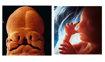
And because it is especially graphic and artistic, I must include Lennart Nilsson’s jaw-dropping intrauterine endoscopic photography, and, you will sigh on learning this, artistically lighted and arranged macro shots of aborted embryonic parts, or, as we pathologists report them, “products of conception,” POC. No dearth of material.
For scientifically accurate depiction of gross but not microscopic embryology, Nilsson’s is the most effective I’ve seen. Not confined to the scientific community, his photographs were featured in the 1960s by a series of famous Life Magazine cover stories and TV’ Nova” movie, “Life’s Greatest Miracle,” from which I learned more, with more awe, than from my med school embryology. For simple primitive awe, and as photographic art an Emmy winner, but of less help to a med student preparing for the final exam today, Nilsson glorified the unseen embryo as Michelangelo glorified the muscled man.
Getting my breath back, and getting back to academic embryology, and electron microscopy (not exactly art photography but in certain ways more informative), the other day I visited my med school book store and thumbed through the embryo text med students are now studying. Sure different from mine. No indeed, embryogenesis is not as I learned it so long ago. Likewise changed, the philosophy of it, notably Evolutionary embryology.
As to new knowledge in embryogenesis, take, for example, intracellular, or primary or non-motile, cellular cilia, as determined by electron microscopy. In my day, the day of light microscopy, cilia were the huge, actively waving external projections that waft an ovum down the Fallopian duct or eject flotsam and jetsam from the lungs or sinuses. Now cilia are so much more, and more minuscule, than that.
The newly-discovered cilia are inside the cell, usually deep inside, and termed “nonmotile” to distinguish them from the familiar moving arms projecting from the cell. But intracellular cilia are anything but immobile. They move, and they move stuff, inside the cell. If DNA, also new since my day (we knew about chromosomes and the laws of genetics but not the DNA molecule), is genetic information, the intra-cellular cilia and integrally related structures, notably the centriole, is the logistics for deploying that information in meiosis and mitosis. Those intracellular cilia and attached and related structures, now seen in as great detail as in a blueprint of a jet engine rotor (they remind me of Gatling guns), are the smart-tugboats or smart-forklifts scurrying around the Amazon warehouse, more like intelligent robots that move and intelligently assemble other intracellular components, large and small, all over the intracellular scene. And they do much more than that.
Confused by the use of the term “cilia” to encompass structures so different in size, function, and location, old students wonder why those tiny streaks inside the cell are even called “cilia.” It is because, I assume, that on an electron microscopic level the intricate internal anatomy of both the large and tiny kinds are so strikingly similar, surprisingly so, which to the enquiring mind triggers the whole business about the philosophy of it all, especially Evolutionary philosophy, which itself has evolved, obviously by mutation, from a philosophy in my day, to science itself, science sovereign over all other science, and, even further, the law of the land.
An eon ago, when I was a premed student with a biology major, I was subjected to Haeckel’s diagrams showing from left to right the development of early absolutely identical embryos of fish, salamander, turtle, chicken, rabbit, and human all progressively burgeoning and bulging and self-sculpting identical brachial clefts and somites in sync, all proving, according to Haeckle’s motto accompanying it, that "ontogeny recapitulates phylogeny." Haeckle’s diagram was a banner waved in the faces of creationists. Thus empowered embryology was one of the flagships of evolution, along with paleontology, geology, anthropology, biology..
An admirer and roughly a contemporary of Darwin, Haeckel was a renowned naturalist, philosopher, physician, professor, a good coiner of words (“phylogeny” is his) and mottos (“Ontogeny recapitulates…” is his), and also a pretty good artist (he himself drew the diagrams), though prone, like artists as a species are, to artistic license, as many of his apologists have, with chagrin, admitted. Likewise for the motto, as Wikipedia forthwith admits, sort of. Quibble not withstanding, Haeckle’s diagram and motto, in my day momentarily convincing, are now as obsolete as an embryonic neural crest in a tattooed computer engineer.
Haeckle and Darwin, and for a few generations their disciples, using only eyeballs and primitive microscopes, dealt with things (embryos, rocks, fossils, and so on) that looked like they could be arranged in a meaningful order, and inferred not illogically they had been generated in sequence and related. Alas, the recent so much more detailed revelations yielded by the so much more sophisticated research technology have yielded philosophical problems.
Just for example, those intracellular cilia, which by early simple electron microscopy looked like only bad halftone graininess from an 1890 newspaper picture or spilled buckshot without function or purpose. Such a pile of refuse would be a big problem for a Creationist whose God had neatly carried off all creation in 6 days with clarity and certainty, of which embryogenesis is a model.
But for Evo no problem. Junk organelles – junk. Junk is what Evo says they are, embarrassingly reminiscent of what Evo had called most DNA -- junk DNA. Easy for Evo to say because junk is exactly what the law nee theory of Evolution predicts and requires. Being a matter of millions and billions of years of experimentation, failed luck, and random trial and error, mostly error, Evolution naturally leaves piles of spent casings or sawdust, rubbish, the sheer bulk of which would come to outweigh what has turned out to be useful, thus proving Evolution. Trained in the law of probability, a true scientist expects most of his lab experiments not pan out, so why should those of Evolution, the truest possible science? The greater the number of things that are useless, the stronger Evo’s case.
The mechanism classically invoked is random mutation of genetic material, thereby generating more complicated molecules from which come, after an eon or two of experimental assembly, more complicated structures, thus transforming a prokaryocyte to a eukaryotye. In real life, in a real lab, even in court, more mutations are disadvantageous than advantageous – sending the process the wrong way. But as in religion faith overcomes, so in Evolution eons do.
To me at least as credible, and a lot more charming, and neater, is Lewis Thomas’s explanation, presented in light-hearted essays and humorous free verse. Lewis Thomas, MD, roughly a contemporary of mine, a renowned naturalist, physician (Harvard grad; dean of Yale Medical School), administrator, longtime editor the New England Journal of Medicine, essayist, poet, and light-hearted Evo apologist, explains: implants. Rather than being the products of eons of experimental mutation, waving cilia are implanted whole spirochetes. Likewise centrioles and mitochondria are implanted animalcules of some sort, he’s not sure but it doesn’t matter, illegal aliens granted amnesty, that decided to pay taxes and be productive citizens. Chuckling, I think that what he mainly has proved is that Evo could have evolved a funny bone, as handy an accouterment to Evo at lectern and blog as cilia in the Fallopian tube. The planet is saddened that the great Funny-boneosaurus has become extinct.
Meanwhile back in the uterus embryogenesis-ontogency has rolled right along as if by a master cyber program, which is a feeble simile for the master divine plan of creation that indeed is in control, the spirit of God moving upon the waters, and a new life has materialized among us. Embryogenesis finished and the product of it having been introduced to the world, that's not the end, that’s just the beginning, yet another beginning. For the next 20 years or even lifetime, not a mere 9 months, the mother, also the father, must, or should, set about training this boy or girl, once just a tuft of cells, to be a functional, productive citizen rather than Evolutionary or social junk, a mature human being, in the image of God, and that task, in a way, is at least as tough as God’s of forming the embryo in the womb. It may turn out to be a medical student.
In The Beginning
Photos by Lennart Nilsson